Cell Dynamics
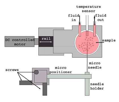 |
Figure 1. Experimental setup to study the mechanical response of the axon to an external force. The force is applied by using a micro needle and moving the sample through a micropositioner. The sample temperature remains constant with the help of a controlled thermal bath.
One of the most outstanding characteristics of the axons is related to its dimension, they possess a typical average radius of 0.5 microns while their length vary from a few dozens of microns to 1 meter, two millions times the typical radius!. From a purely mechanical point of view, an axon has similar characteristics to a string, however its internal structures is composed by tubulin molecules, microtubules and molecular motors among other complex structures. The interactions between the different filaments and the molecular motors play an important role in determining the mechanical properties of the axons.
Our main objective is to develop new techniques and materials to be used as test in biological systems. We have applied our expertise on the physics of crumpling surfaces to develop and characterize thin substrata. These substrata, of the order less of one micron thickness and obtained by the vulcanization of a layer of silicon fluid, are well adapted to measure forces applied by living cells during their motion. Such forces, of the order of a nanonewton, when applied on our substrate produce well-defined wrinkles. We have developed interferential methods to accurately measure the wrinkles topology which in turn, by the use of elasticity equations, allows us to measure cell forces. Thus, cell motion strategies and cell adhesion can be accurately investigated. Our preliminary results include the observation of well defined wrinkles produced by fibroblasts and the characterization of thin substrata by micromechanical methods. We have achieved to observe wrinkles produced by fibroblasts in a new apparatus that integrates the interferometer, the temperature control and the cellular medium. Recently we have achieved to measure the thickness of our substrate and the residual tension acting on it. Both parameters are important to obtain quantitative information on the forces acting on the membrane by the action of the cell.
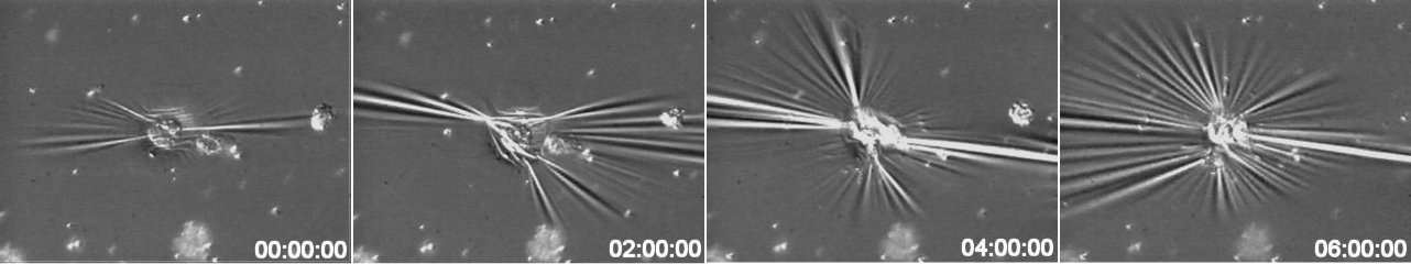
Time sequence of a contracting cell. Radial wrinkles are clearly visible under DIC microscopy.